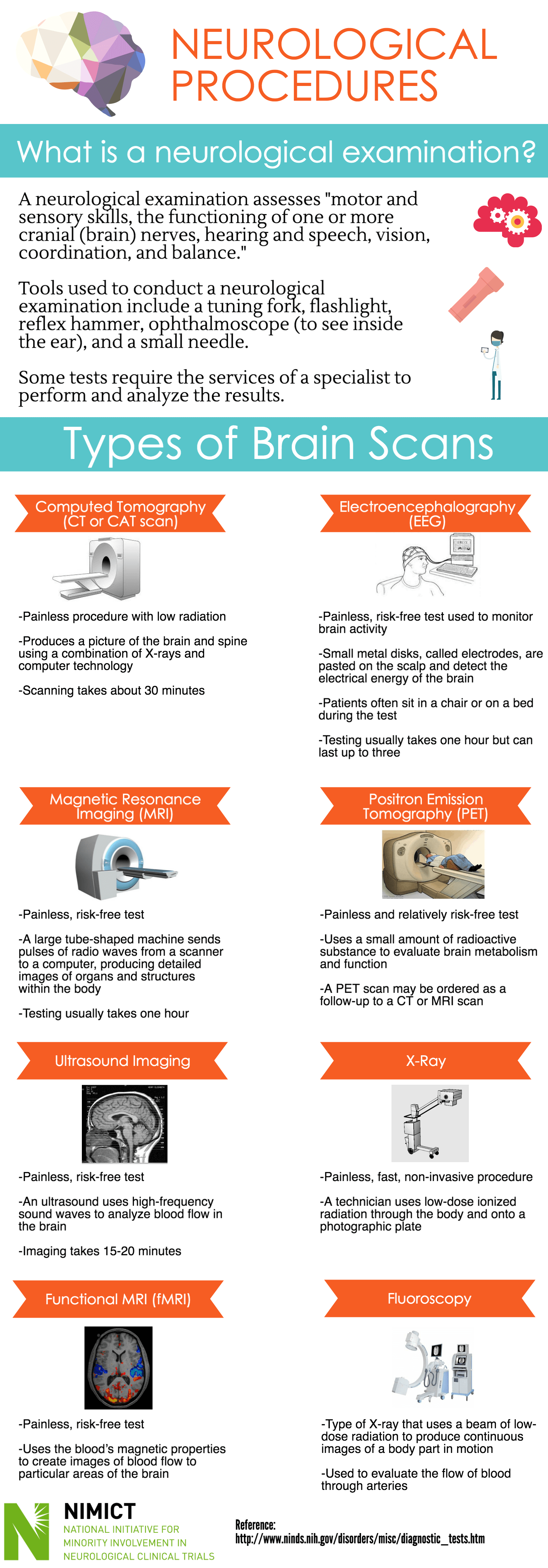
Neurological Procedures
NIMICT Original

Neurological Procedures
What is a neurological examination?
-A neurological examination assesses "motor and sensory skills, the functioning of one or more cranial (brain) nerves, hearing and speech, vision, coordination, and balance."
-Tools used to conduct a neurological examination include a tuning fork, flashlight, reflex hammer, ophthalmoscope (to see inside the ear), and a small needle.
-Some tests require the services of a specialist to perform and analyze the results.
Types of Brain Scans
Computed Tomography (CT or CAT scan)
-Painless procedure with low radiation
-Produces a picture of the brain and spine using a combination of X-rays and computer technology
-Scanning takes about 30 minutes
Electroencephalography (EEG)
-Painless, risk-free test used to monitor brain activity
-Small metal disks, called electrodes, are pasted on the scalp and detect the electrical energy of the brain
-Patients often sit in a chair or on a bed during the test
-Testing usually takes one hour but can last up to three
Magnetic Resonance Imaging (MRI)
-Painless, risk-free test
-A large tube-shaped machine sends pulses of radio waves from a scanner to a computer, producing detailed images of organs and structures within the body
-Testing usually takes one hour
Positron Emission Tomography (PET)
-Painless and relatively risk-free test
-Uses a small amount of radioactive substance to evaluate brain metabolism and function
-A PET scan may be ordered as a follow-up to a CT or MRI scan
Ultrasound Imaging
-Painless, risk-free test
-An ultrasound uses high-frequency sound waves to analyze blood flow in the brain (imaging takes 15-20 minutes)
X-Ray
-Painless, fast, non-invasive procedure
-A technician uses low-dose ionized radiation through the body and onto a photographic plate
Functional MRI (fMRI)
-Painless, risk-free test
-Uses the blood’s magnetic properties to create images of blood flow to particular areas of the brain
Fluoroscopy
-Type of X-ray that uses a beam of low dose radiation to produce continuous images of a body part in motion
-Used to evaluate the flow of blood through arteries